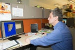
All the Cores at the Center for Craniofacial Regeneration provide service to researchers from academic and research institutions across the United States, as well as those at the University of Pittsburgh. Among the state-of-the-art resources and equipment available are microCT scanners as well as scanners and software for histology and numerous apparatus for specimen preparation and mechanical testing.
Microcomputed Tomography
Director: Konstantinos Verdelis, DDS, PhD
Equipment: Scanco VivaCT 40; Scanco µCT50
The CCR microCT Core suite occupies 300 square feet on the fourth floor of the Salk Pavilion building. This core uses an in vivo Scanco VivaCT 40, Scanco Medical, scanner based at the Bridge Side Point II building, just over a mile from Salk Pavillion. This equipment is capable of intermediate resolutions (10.5-40µm voxel resolution) imaging of live or harvested tissues. The microCT Core also includes a newly acquired Scanco µCT50, Scanco Medical high resolution (0.4-30µm voxel resolution) ex vivo scanner. The Scanco µCT50 scanner is capable of imaging wide objects (up to 10cm extended field of view) with a wide range of densities (up to 90Kvp beam energy range for implant imaging). It has a high throughput capability through the use of an autoloader and pre-programming of individual sample runs. Applications range from imaging of hard tissues to biomaterials and soft tissues (like vasculature, embryonic tissue and joints and ligaments). The CCR microCT Core facility employs a dedicated technical specialist and has processing and user-training areas for the use of Scanco, Bruker-Skyscan and Mimics 3D morphometry software on dedicated HP Raid tower and Dell workstations.
For your reference, please see our microCT fees, below:
Academic (University of Pittsburgh)
Imaging - $40.00/hr
Analysis - $40.00/hr
Academic (external) & Non-profit
Imaging - $65.00/hr
Analysis - $65.00/hr
Commercial
Imaging - $100/hr
Analysis - $100/hr
Histology
Director: Charles S. Sfeir, DDS, PhD
Equipment: Automatic Tissue Processor; Embedder; Stainer; Microtomes; Microscopes including: diffraction, brightfield, dissecting and fluorescent microscopes
The CCR histology suite occupies 450 square feet on the fifth floor of the Salk Pavilion building. It is dedicated to the histology of soft tissues, decalcified and undecalcified hard tissues, interfaces of tissues with biomaterials such as implanted metal devices, histomorphometry, as well as immunohistochemistry and in situ hybridization analysis. A dedicated technical specialist is responsible for the operation of this core, including training and supervision of users. A range of microtomes (rotary, heavy duty controlled-speed and cryostats) is available to match the particular specimens and purpose of analysis. The suite houses an automatic tissue processor, embedder and stainer equipment, which make high sample turnover possible. The instruments used include diffraction, brightfield and fluorescent microscopes; ones equipped with cameras, as well as several dissecting microscopes. In addition to using widely established techniques for soft tissues, the core has developed and uses methodologies to perform advanced histological and histomorphometric assessments of mineralized tissues (based on PMMA and like proprietary resins, Technovit processing). Histomorphometry is performed using high quality scanners and specialized software (such as that created by Bioquant) on dedicated workstations.
Biomechanics
Director: Alejandro Almarza, PhD
Equipment: Dual Column Instron testing system; Single Column MTS testing system; Buehler Microhardness Tester
The Biomechanics Core is housed within the Center for Craniofacial Regeneration on the fourth floor of Salk Pavilion with areas for specimen preparation and mechanical testing. The core uses a dual column Instron material testing apparatus, a single column MTS testing apparatus and a Buehler microhardness tester. Mechanical properties of hard and soft tissues and bone are characterized by a range of tests, such as unconfined compression, uniaxial tension, 3 and 4 point bending, and puncture. Multiple load cells on instruments provide a range of mNewton to kNewton for testing, strain gauges and video tracking, which are used to validate deformations. The systems are also equipped to test new materials, including metal alloys.


